
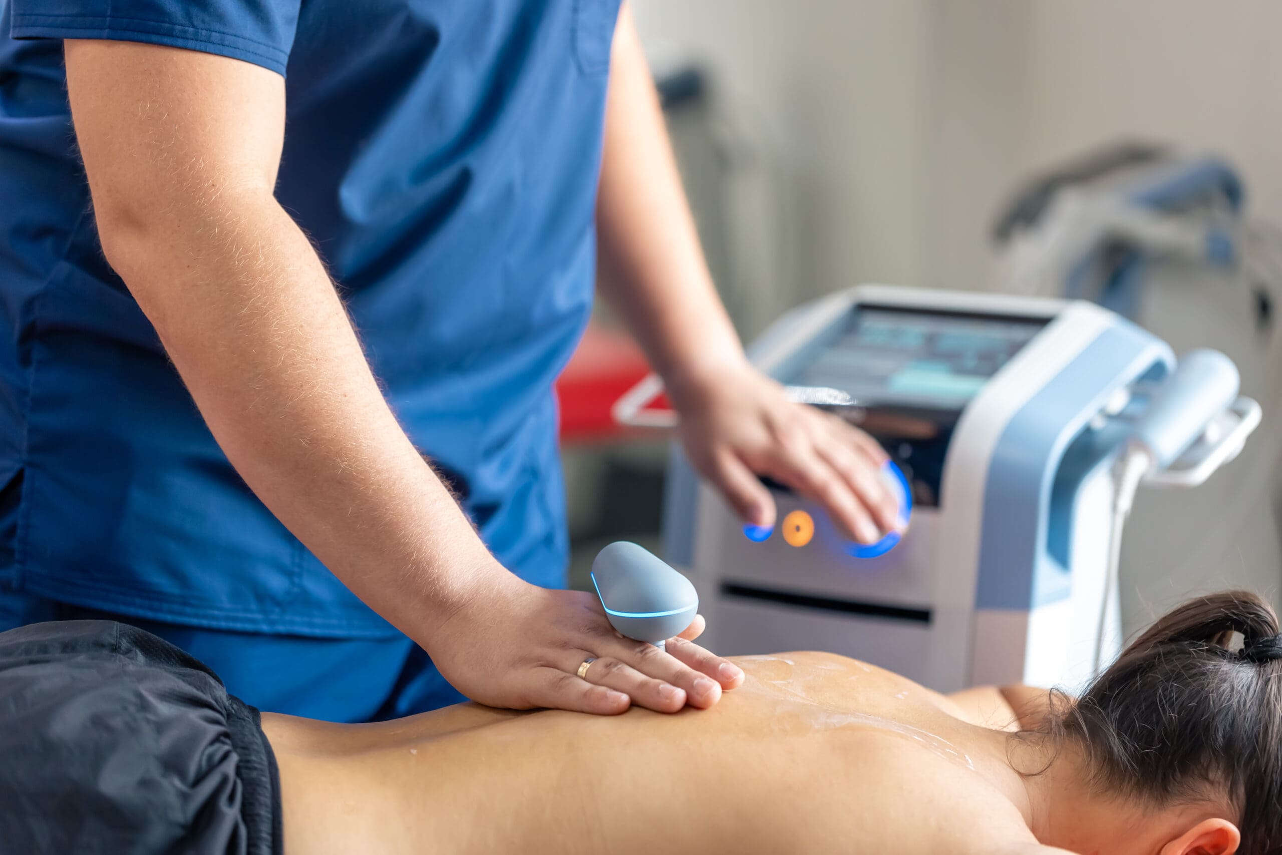
Discover how ultrasound therapy can lead to better outcomes in functional recovery and musculoskeletal pain management.
Table of Contents
Abstract
Musculoskeletal (MSK) ultrasound has revolutionized the way we, as clinicians, visualize, diagnose, and treat a wide array of soft-tissue conditions. This technology offers a real-time, dynamic view of anatomy that is simply unparalleled by static imaging like X-rays or even MRIs in many contexts. In this educational post, I will take you on a journey through the fundamental principles of MSK ultrasound, translating the complex world of sonographic patterns into an easy-to-understand narrative. We will explore how different tissues—tendons, muscles, cartilage, ligaments, and nerves—appear on the screen and what these appearances signify. I will share insights from leading researchers and evidence-based methods, along with my own clinical observations, to demonstrate the power of this diagnostic tool. Furthermore, we will delve into the practical skills of probe handling, the importance of avoiding common pitfalls like anisotropy, and how to set yourself up for success in both diagnostic and interventional procedures. Finally, I will explain how these advanced imaging techniques integrate seamlessly into a comprehensive chiropractic care model, enhancing our ability to provide precise, effective, and patient-centered treatment.
Understanding the Language of Ultrasound: Interpreting Tissue Echogenicity
The core of ultrasound imaging lies in understanding echogenicity, which is essentially how bright or dark a tissue appears on the screen. This is determined by how the ultrasound waves reflect off the tissue and return to the probe. Tissues that reflect a lot of sound waves are called hyperechoic and appear bright white, while tissues that reflect few waves are hypoechoic and appear dark or black. Tissues with similar brightness to their surroundings are described as isoechoic.
Recognizing the specific patterns associated with different tissues is a critical skill. It’s like learning to read a new language—the language of the body’s internal structures. Let’s break down the characteristic appearances of key musculoskeletal tissues.
Tendons: The Fibrillar Pattern
When we look at a healthy tendon, we expect to see a distinct hyperechoic (bright) fibrillar pattern. This looks like a series of tightly packed, parallel white stripes. A perfect example is the patellar tendon. When viewed in a long-axis orientation (in line with the tendon fibers), you can clearly see this organized, fiber-like structure.
- Hyperechoic: Appears bright white on the ultrasound image.
- Fibrillar Pattern: A distinct, striped appearance representing organized collagen fibers.
- Anatomical Context: In the image of the patellar tendon, you can see the tendon connecting the patella (kneecap) on the left to the tibia (shin bone) on the right. Below the tendon, the wavy, darker structure is the infrapatellar fat pad.
This pattern recognition is fundamental. When this organized, bright, striped pattern is disrupted—becoming darker, thicker, or disorganized—it can indicate pathology such as tendinosis or a tear.
Muscles: A Feather-Like Appearance
Normal muscle tissue presents a more complex, mixed-echogenicity pattern. It is generally hypoechoic (darker) compared to the stark white appearance of bone. However, within the muscle belly, you’ll see hyperechoic (brighter) strands of fibro-adipose septa and perimysium, which are connective tissues that wrap around muscle fiber bundles. This gives the muscle its characteristic “feather-like” or “starry night” appearance in a textbook view.
- Hypoechoic Background: The muscle fibers themselves are dark.
- Hyperechoic Strands: The connective tissue within the muscle appears as bright white lines.
- Bone: Appears as a very bright, continuous white line with a dark “acoustic shadow” beneath it, as the sound waves cannot penetrate it.
From a single image, I can identify a muscle overlying a bone, such as the humerus. Still, I would need to scan along its course to definitively identify it as the biceps or deltoid. This ability to trace structures is a unique advantage of ultrasound.
Cartilage: Differentiating Hyaline and Fibrocartilage
In ultrasound, cartilage is not a single entity; we must differentiate between hyaline cartilage and fibrocartilage.
- Hyaline Cartilage: This is the smooth, articular cartilage that lines the surfaces of joints. On ultrasound, it appears as a distinct hypoechoic (dark) line sitting directly on top of the bright white cortical bone. On the shoulder, for instance, you can see this smooth black stripe covering the humeral head.
- Fibrocartilage: This type of cartilage, found in structures like the glenoid labrum in the shoulder or the meniscus in the knee, is much brighter. It appears hyperechoic, distinguishing it from the adjacent hyaline cartilage.
This distinction is clinically vital. Damage to the dark hyaline cartilage can indicate osteoarthritis, while a tear in the bright fibrocartilage of the labrum can be a source of shoulder instability and pain.
The Dynamic Duo: Ligaments and Nerves
Ligaments: Densely Packed Fibers
Ligaments look very similar to tendons, displaying a hyperechoic, striated, fibrillar pattern. However, there are key differences:
- Anatomical Connection: The most definitive way to identify a ligament is by its connections. Ligaments connect bone to bone. If you trace a structure and it attaches from one bony prominence to another, it’s a ligament. If it attaches muscle to bone, it’s a tendon.
- Fiber Density: Ligament fibers are typically more densely packed and more compact than tendon fibers, giving them an even more tightly woven appearance on ultrasound.
The real power of ultrasound in assessing ligaments is its dynamic capability. I can perform a stress test in real-time. For example, when evaluating the Medial Collateral Ligament (MCL) of the knee, I can apply a valgus stress (pushing the knee inward) while imaging the ligament. If the ligament is torn, I will see the tissue gap open on the screen. This allows me to grade the sprain (Grade 1, 2, or 3) right at the point of care, providing immediate, actionable diagnostic information. This is something a static MRI cannot offer.
Nerves: The Honeycomb Structure
Nerves have a fascinating, distinctive appearance that makes them identifiable. In a cross-sectional (short-axis) view, a nerve looks like a honeycomb.
- Fascicles: The nerve fascicles (bundles of nerve fibers) are hypoechoic (dark circles).
- Epineurium: The connective tissue sheath surrounding the fascicles, known as the epineurium, is hyperechoic (bright white lines).
This creates a mixed-appearance, honeycomb pattern that is a hallmark of nerve tissue. In the long-axis view, it appears as parallel dark bands (the fascicles) separated by bright lines (the epineurium), but the honeycomb view is often more distinct.
A pro-tip for finding nerves: scan. Your eye is naturally drawn to patterns that move and change. As you scan rapidly across a region like the forearm, the nerve’s unique honeycomb texture will “pop” out from the more linear and uniform appearance of the surrounding muscles and tendons. The median nerve in the carpal tunnel is a classic example in which its echogenicity is distinctly different from that of the adjacent flexor tendons, making it a perfect spot to practice this technique.
Avoiding A Common Pitfall: The Challenge of Anisotropy
One of the most important concepts to master in MSK ultrasound is anisotropy. This is an artifact, not a true pathological finding, that occurs when the ultrasound beam is not perfectly perpendicular to the tissue being imaged, particularly in highly organized structures such as tendons.
When the beam hits the tendon at an angle, the sound waves reflect away from the probe instead of back to it. This lack of return signal causes the tendon to appear artifactually hypoechoic (dark), mimicking a tear or tendinosis.
- The Rule: If you see a dark area in a tendon, the first thing you must do is adjust your probe by rocking it back and forth (a technique called “heel-toeing”) to ensure you are perfectly perpendicular to the fibers at that spot.
- True Pathology vs. Anisotropy:
- If the dark spot disappears when you adjust the probe, it was anisotropy.
- If the dark spot remains dark no matter how you angle the probe, it is more likely to be true pathology.
I always follow the surgical principle: “one view is no view”. If I suspect a tear, I must prove it. I will image it from multiple angles along both the long and short axes and perform dynamic testing. For a suspected rotator cuff tear, I would have the patient resist abduction. If the dark area gaps open with muscle contraction, that confirms a tear. Relying on a single image can lead to a misdiagnosis. You must use all the tools in your toolbox to convince yourself of the finding.
Mastering the Craft: Probe Handling for Diagnosis and Intervention
How you hold the ultrasound probe is not a trivial matter; it is the foundation of acquiring good images and performing procedures safely and effectively.
The Tripod Technique
For diagnostic scanning, we use the tripod technique. This involves holding the probe like a pencil and using your pinky, ring, and/or middle finger to brace your hand against the patient’s skin. This creates a stable base, allowing for fine, controlled movements. You must have physical contact with the patient; holding the probe by its tail or cord gives you no control and results in poor-quality images.
My clinical observation is that many practitioners are trained to wrap their entire hand around the probe for stability. While this is indeed stable for diagnostic scanning, it becomes a major obstacle for interventional procedures. Your own fingers get in the way of where the needle needs to go, compromising sterility and access.
Adapting for Interventional Procedures
When I prepare for an injection, I modify my grip. I hold the probe by its edges, which keeps my fingers clear of the sterile field and the needle path. This allows me to maintain a perfect view of my target while having an unobstructed path for my needle.
The goal is to set yourself up for success.
- Find the Target: Use gross scanning movements to find the general area, then fine-tune your position to get the best possible image of your target structure.
- Stay Perpendicular: Make every effort to keep your probe perpendicular to the target. This minimizes anisotropy and gives you the clearest view.
- Plan Your Trajectory: By keeping the target directly beneath the probe’s center, you simplify your needle path. You can plan a straightforward trajectory instead of calculating complex angles, which dramatically increases the likelihood of a successful and efficient procedure.
My approach is to orient the probe to the patient’s anatomy, not to a fixed convention on the screen. I set the screen so that the patient’s right is on the right side of the screen and their left is on the left. This eliminates the mental gymnastics of having to reverse the image in my head while maneuvering a needle in real-time. It makes the procedure faster, safer, and more intuitive.
Integrating Ultrasound into Chiropractic and Functional Medicine
As an integrative practitioner, I view the body as a connected system. Musculoskeletal ultrasound fits perfectly into this holistic framework. It’s not just a diagnostic “flashlight”; it’s a tool that enhances our therapeutic precision.
In my practice, chiropractic care focuses on restoring proper biomechanics and nervous system function through adjustments, soft tissue work, and rehabilitative exercises. Ultrasound provides an inside look at the structural integrity of the tissues we are treating.
- Precision Diagnosis: Before beginning treatment for shoulder pain, I can use an ultrasound to differentiate between a rotator cuff tear, biceps tendinopathy, bursitis, or labral pathology. This allows me to tailor the treatment plan specifically to the injured tissue. A chiropractic adjustment might be central for restoring joint mobility, but knowing there’s a partial tendon tear means I will also incorporate specific regenerative therapies and avoid movements that could worsen the injury.
- Tracking Healing: Ultrasound allows me to objectively monitor a patient’s progress. I can visually track the healing of a tendon tear or the reduction of inflammation in a bursa over the course of treatment. This provides valuable feedback for both the patient and me, confirming that our integrative approach—combining chiropractic adjustments, functional nutrition to support tissue repair, and targeted rehabilitation—is working.
- Guided Interventions: For cases requiring more direct intervention, ultrasound guidance is the gold standard. When performing regenerative injections such as platelet-rich plasma (PRP) or prolotherapy, I can guide the needle with millimeter-level accuracy to the exact site of injury. This ensures that the healing agents are delivered precisely where they are needed most, maximizing the therapeutic benefit and improving patient outcomes.
Ultimately, MSK ultrasound empowers us to move beyond assumption and into a state of clinical certainty. It bridges the gap between a physical exam and a definitive diagnosis, allowing for highly specific, evidence-based care plans that integrate the best of chiropractic, functional medicine, and modern interventional techniques. It is an amazing and indispensable tool in my mission to provide the highest level of care to my patients.
References
- Jacobson, J. A. (2017). Fundamentals of musculoskeletal ultrasound (3rd ed.). Elsevier.
- Finnoff, J. T., & Hall, M. M. (Eds.). (2021). Musculoskeletal ultrasound in sports medicine. Springer.
- Bianchi, S., & Martinoli, C. (2007). Ultrasound of the musculoskeletal system. Springer-Verlag.
SEO Tags: Musculoskeletal Ultrasound, MSK Ultrasound, Integrative Chiropractic, Dr. Alexander Jimenez, Echogenicity, Anisotropy, Tendon Imaging, Nerve Ultrasound, Ligament Tear, Ultrasound Guided Injection, Functional Medicine, Chiropractic Care, Point of Care Ultrasound, Rotator Cuff Tear, Carpal Tunnel Syndrome, Diagnostic Ultrasound, Probe Handling, Sports Medicine, Regenerative Medicine
Post Disclaimer
Professional Scope of Practice *
The information herein on "Ultrasound Therapy for Musculoskeletal Pain Management" is not intended to replace a one-on-one relationship with a qualified health care professional or licensed physician and is not medical advice. We encourage you to make healthcare decisions based on your research and partnership with a qualified healthcare professional.
Blog Information & Scope Discussions
Welcome to El Paso's Premier Wellness, Personal Injury Care Clinic & Wellness Blog, where Dr. Alex Jimenez, DC, FNP-C, a Multi-State board-certified Family Practice Nurse Practitioner (FNP-BC) and Chiropractor (DC), presents insights on how our multidisciplinary team is dedicated to holistic healing and personalized care. Our practice aligns with evidence-based treatment protocols inspired by integrative medicine principles, similar to those on this site and our family practice-based chiromed.com site, and focuses on restoring health naturally for patients of all ages.
Our areas of multidisciplinary practice include Wellness & Nutrition, Chronic Pain, Personal Injury, Auto Accident Care, Work Injuries, Back Injury, Low Back Pain, Neck Pain, Migraine Headaches, Sports Injuries, Severe Sciatica, Scoliosis, Complex Herniated Discs, Fibromyalgia, Chronic Pain, Complex Injuries, Stress Management, Functional Medicine Treatments, and in-scope care protocols.
Our information scope is multidisciplinary, focusing on musculoskeletal and physical medicine, wellness, contributing etiological viscerosomatic disturbances within clinical presentations, associated somato-visceral reflex clinical dynamics, subluxation complexes, sensitive health issues, and functional medicine articles, topics, and discussions.
We provide and present clinical collaboration with specialists from various disciplines. Each specialist is governed by their professional scope of practice and their jurisdiction of licensure. We use functional health & wellness protocols to treat and support care for musculoskeletal injuries or disorders.
Our videos, posts, topics, and insights address clinical matters and issues that are directly or indirectly related to our clinical scope of practice.
Our office has made a reasonable effort to provide supportive citations and has identified relevant research studies that support our posts. We provide copies of supporting research studies upon request to regulatory boards and the public.
We understand that we cover matters that require an additional explanation of how they may assist in a particular care plan or treatment protocol; therefore, to discuss the subject matter above further, please feel free to ask Dr. Alex Jimenez, DC, APRN, FNP-BC, or contact us at 915-850-0900.
We are here to help you and your family.
Blessings
Dr. Alex Jimenez DC, MSACP, APRN, FNP-BC*, CCST, IFMCP, CFMP, ATN
email: [email protected]
Multidisciplinary Licensing & Board Certifications:
Licensed as a Doctor of Chiropractic (DC) in Texas & New Mexico*
Texas DC License #: TX5807, Verified: TX5807
New Mexico DC License #: NM-DC2182, Verified: NM-DC2182
Multi-State Advanced Practice Registered Nurse (APRN*) in Texas & Multi-States
Multi-state Compact APRN License by Endorsement (42 States)
Texas APRN License #: 1191402, Verified: 1191402 *
Florida APRN License #: 11043890, Verified: APRN11043890 *
Colorado License #: C-APN.0105610-C-NP, Verified: C-APN.0105610-C-NP
New York License #: N25929, Verified N25929
License Verification Link: Nursys License Verifier
* Prescriptive Authority Authorized
ANCC FNP-BC: Board Certified Nurse Practitioner*
Compact Status: Multi-State License: Authorized to Practice in 40 States*
Graduate with Honors: ICHS: MSN-FNP (Family Nurse Practitioner Program)
Degree Granted. Master's in Family Practice MSN Diploma (Cum Laude)
Dr. Alex Jimenez, DC, APRN, FNP-BC*, CFMP, IFMCP, ATN, CCST
(Board Certified: Family Practice Nurse Practitioner—Multistate)*
(Licensed Nurse Practitioner & Chiropractor - Multistate)*
Clinical Director
Digital Business Card
Dr. Maria Cardenas, MD
(Board Certified: Internal Medicine)
(Licensed Medical Doctor)
Medical Director, Clinical Director & Collaborative Physician
NPI # 1164426749
MD License #: J2933
Licenses and Board Certifications:
MD: Medical Doctor
DC: Doctor of Chiropractic
APRNP: Advanced Practice Registered Nurse
FNP-BC: Family Practice Specialization (Multi-State Board Certified)
RN: Registered Nurse (Multi-State Compact License)
CFMP: Certified Functional Medicine Provider
MSN-FNP: Master of Science in Family Practice Medicine
MSACP: Master of Science in Advanced Clinical Practice
IFMCP: Institute of Functional Medicine
CCST: Certified Chiropractic Spinal Trauma
ATN: Advanced Translational Neutrogenomics
Memberships & Associations:
TCA: Texas Chiropractic Association: Member ID: 104311
AANP: American Association of Nurse Practitioners: Member ID: 2198960
ANA: American Nurse Association: Member ID: 06458222 (District TX01)
TNA: Texas Nurse Association: Member ID: 06458222
NPI: 1205907805
| Primary Taxonomy | Selected Taxonomy | State | License Number |
|---|---|---|---|
| No | 111N00000X - Chiropractor | NM | DC2182 |
| Yes | 111N00000X - Chiropractor | TX | DC5807 |
| Yes | 363LF0000X - Nurse Practitioner - Family | TX | 1191402 |
| Yes | 363LF0000X - Nurse Practitioner - Family | FL | 11043890 |
| Yes | 363LF0000X - Nurse Practitioner - Family | CO | C-APN.0105610-C-NP |
| Yes | 363LF0000X - Nurse Practitioner - Family | NY | N25929 |
Dr. Alex Jimenez, DC, APRN, FNP-BC*, CFMP, IFMCP, ATN, CCST
(Board Certified: Family Practice Nurse Practitioner—Multistate)*
(Licensed Nurse Practitioner & Chiropractor - Multistate)*
Clinical Director
Digital Business Card
Dr. Maria Cardenas, MD
(Board Certified: Internal Medicine)*
(Licensed Medical Doctor)*
Medical Director, Clinical Director & Collaborative Physician
NPI # 1164426749
MD License #: J2933


 Again, We Welcome You.
Comments are closed.