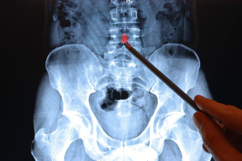
Low back pain is one of the most common ailments for people visiting a doctor or an urgent care clinic. When the back pain becomes intense it can get you thinking something is seriously wrong with your back. The doctor might offer an x-ray or MRI scan to put your concerns at ease.
Fortunately, most cases for low back pain even acute pain improve within days or a few weeks. Most cases are remedied with chiropractic, physical therapy, heat/ice therapy and rest. And a lot of these cases do not require any form of spinal imaging. However, there are those reasons when X-ray, MRI, CT scans are necessary to figure out what’s going on.
- Strained muscle
- Sprained ligament
- Poor posture
These are typical causes of low back pain that can be painful and limit activities.

Table of Contents
Back Pain Lasting Longer Than 2/3 Weeks
Subacute pain lasts between 4 and 12 weeks while chronic back pain lasts 3 months or longer. These are not indications of a serious low spinal condition.
Less than 1% of people with low back pain are diagnosed with a condition that may require spine surgery like:
- Cauda equina syndrome
- Spinal infection
- Metastatic spinal cancer
X-rays or MRI’s for Diagnosing Low Back Pain
Doctors may recommend an x-ray or MRI if the low back pain was from a traumatic injury, like a:
- Slip
- Fall
- Automobile accident
Other potential causes of low back pain may warrant medical imaging immediately or later on.
The diagnostic process starts with the evaluation of the low back symptoms and how they relate to what was found during the:
- Physical exam
- Neurological exam
- Medical history
A doctor utilizes these results to figure out whether the need for spinal imaging is necessary, along with the type of imaging test, x-ray or MRI and the timing to confirm a diagnosis.
A Low Back X-Ray/MRI
X-ray spinal imaging is best at detecting bony structural problems but not so great with soft tissue injuries. There are X-ray series that may be performed to diagnose vertebral compression fractures like.
- Anterior
- Posterior
- Lateral views
MRI is a radiation-free test. MRI’s create 3-D anatomical views of the spinal bones and soft tissues. A contrast dye like gadolinium is used to enhance and improve the quality of the images. The contrast is injected through an intravenous line in your hand or arm before or during the test. An MRI can evaluate neurological symptoms, like radiating pain or pain that develops after being diagnosed with cancer.
Symptoms, Co-existing Medical Diagnoses, and Conditions that may Require Spine Imaging
Neurological symptoms
- Low back pain that radiates, fans out or downward into the buttocks, legs, and feet
- Abnormal reflexes in the lower body can indicate nerve disruption
- Numbness, tingling, and possibly weakness develop
- Inability to lift your foot aka foot drop
Co-existing medical diagnoses and conditions
- Cancer
- Diabetes
- Fever
- Osteoporosis
- Previous spinal fracture
- Spine surgery
- Recent infection
- Immunosuppressant medication use
- Corticosteroid medication
- Weight loss
X-ray Radiation Exposure
When undergoing an x-ray, the radiation not absorbed by the body creates the image. The radiation dose is the same amount every time you undergo an x-ray. Radiation to your entire body is measured through the millisievert (mSv) also known as the effective dose.
The effective dose helps a doctor measure the risk for possible side effects of radiographic imaging:
- CT scans use radiation as well
- Certain body tissues and organs in the lower back are sensitive to radiation exposure like the reproductive organs.
MRI Radiation-Free Why Not Just Use This Test All The Time
MRI’s cannot be used on all patients because of its powerful magnet technology. Pregnant women or individuals that have metal inside their body like a spinal cord stimulator, heart pacemaker, etc cannot be scanned with an MRI.
MRI testing is also expensive, doctors do not want to prescribe unnecessary tests that increase costs. Or because of the fine detail that MRI’s provide, sometimes a spinal issue can look serious but is not.
Example: An MRI of the lower back reveals a herniated disc in a patient that has no back/leg pain or other symptoms.
This is why doctors bring all of their findings like the symptoms, physical exam, and medical history to confirm a diagnosis and then create the custom treatment plan.
Imaging Test Takeaways
If low back pain begins to take its toll, listen to what the doctor recommends. They might not order a lumbar x-ray or MRI immediately but remember the aforementioned issues like neurological symptoms and co-existing medical conditions. But these tests do help in discovering the cause or causes of the pain. Remember this is to help get patients to their optimal health and pain-free.
How to eliminate Back Pain naturally | Foot Levelers | El Paso, Tx
NCBI Resources
Imaging diagnostics is an essential element in the evaluation of spine trauma. The rapid evolution of imaging technology has tremendously changed the assessment and treatment of spine injuries. Imaging diagnostics utilizing CT and MRI, among others, are helpful in acute and chronic settings. Spinal cord and soft-tissue injuries are best evaluated by magnetic resonance imaging, or MRI, whereas computed tomography scanning or CT scans, best evaluate spinal trauma or spine fracture.
Post Disclaimer
Professional Scope of Practice *
The information herein on "Why Do I Need an X-ray or MRI for Lower Back Pain El Paso, TX?" is not intended to replace a one-on-one relationship with a qualified health care professional or licensed physician and is not medical advice. We encourage you to make healthcare decisions based on your research and partnership with a qualified healthcare professional.
Blog Information & Scope Discussions
Welcome to El Paso's Premier Wellness, Personal Injury Care Clinic & Wellness Blog, where Dr. Alex Jimenez, DC, FNP-C, a Multi-State board-certified Family Practice Nurse Practitioner (FNP-BC) and Chiropractor (DC), presents insights on how our multidisciplinary team is dedicated to holistic healing and personalized care. Our practice aligns with evidence-based treatment protocols inspired by integrative medicine principles, similar to those on this site and our family practice-based chiromed.com site, and focuses on restoring health naturally for patients of all ages.
Our areas of multidisciplinary practice include Wellness & Nutrition, Chronic Pain, Personal Injury, Auto Accident Care, Work Injuries, Back Injury, Low Back Pain, Neck Pain, Migraine Headaches, Sports Injuries, Severe Sciatica, Scoliosis, Complex Herniated Discs, Fibromyalgia, Chronic Pain, Complex Injuries, Stress Management, Functional Medicine Treatments, and in-scope care protocols.
Our information scope is multidisciplinary, focusing on musculoskeletal and physical medicine, wellness, contributing etiological viscerosomatic disturbances within clinical presentations, associated somato-visceral reflex clinical dynamics, subluxation complexes, sensitive health issues, and functional medicine articles, topics, and discussions.
We provide and present clinical collaboration with specialists from various disciplines. Each specialist is governed by their professional scope of practice and their jurisdiction of licensure. We use functional health & wellness protocols to treat and support care for musculoskeletal injuries or disorders.
Our videos, posts, topics, and insights address clinical matters and issues that are directly or indirectly related to our clinical scope of practice.
Our office has made a reasonable effort to provide supportive citations and has identified relevant research studies that support our posts. We provide copies of supporting research studies upon request to regulatory boards and the public.
We understand that we cover matters that require an additional explanation of how they may assist in a particular care plan or treatment protocol; therefore, to discuss the subject matter above further, please feel free to ask Dr. Alex Jimenez, DC, APRN, FNP-BC, or contact us at 915-850-0900.
We are here to help you and your family.
Blessings
Dr. Alex Jimenez DC, MSACP, APRN, FNP-BC*, CCST, IFMCP, CFMP, ATN
email: [email protected]
Multidisciplinary Licensing & Board Certifications:
Licensed as a Doctor of Chiropractic (DC) in Texas & New Mexico*
Texas DC License #: TX5807, Verified: TX5807
New Mexico DC License #: NM-DC2182, Verified: NM-DC2182
Multi-State Advanced Practice Registered Nurse (APRN*) in Texas & Multi-States
Multi-state Compact APRN License by Endorsement (42 States)
Texas APRN License #: 1191402, Verified: 1191402 *
Florida APRN License #: 11043890, Verified: APRN11043890 *
Colorado License #: C-APN.0105610-C-NP, Verified: C-APN.0105610-C-NP
New York License #: N25929, Verified N25929
License Verification Link: Nursys License Verifier
* Prescriptive Authority Authorized
ANCC FNP-BC: Board Certified Nurse Practitioner*
Compact Status: Multi-State License: Authorized to Practice in 40 States*
Graduate with Honors: ICHS: MSN-FNP (Family Nurse Practitioner Program)
Degree Granted. Master's in Family Practice MSN Diploma (Cum Laude)
Dr. Alex Jimenez, DC, APRN, FNP-BC*, CFMP, IFMCP, ATN, CCST
My Digital Business Card
Licenses and Board Certifications:
DC: Doctor of Chiropractic
APRNP: Advanced Practice Registered Nurse
FNP-BC: Family Practice Specialization (Multi-State Board Certified)
RN: Registered Nurse (Multi-State Compact License)
CFMP: Certified Functional Medicine Provider
MSN-FNP: Master of Science in Family Practice Medicine
MSACP: Master of Science in Advanced Clinical Practice
IFMCP: Institute of Functional Medicine
CCST: Certified Chiropractic Spinal Trauma
ATN: Advanced Translational Neutrogenomics
Memberships & Associations:
TCA: Texas Chiropractic Association: Member ID: 104311
AANP: American Association of Nurse Practitioners: Member ID: 2198960
ANA: American Nurse Association: Member ID: 06458222 (District TX01)
TNA: Texas Nurse Association: Member ID: 06458222
NPI: 1205907805
| Primary Taxonomy | Selected Taxonomy | State | License Number |
|---|---|---|---|
| No | 111N00000X - Chiropractor | NM | DC2182 |
| Yes | 111N00000X - Chiropractor | TX | DC5807 |
| Yes | 363LF0000X - Nurse Practitioner - Family | TX | 1191402 |
| Yes | 363LF0000X - Nurse Practitioner - Family | FL | 11043890 |
| Yes | 363LF0000X - Nurse Practitioner - Family | CO | C-APN.0105610-C-NP |
| Yes | 363LF0000X - Nurse Practitioner - Family | NY | N25929 |
Dr. Alex Jimenez, DC, APRN, FNP-BC*, CFMP, IFMCP, ATN, CCST
My Digital Business Card


 Again, We Welcome You.
Comments are closed.